Categories
Change Password!
Reset Password!


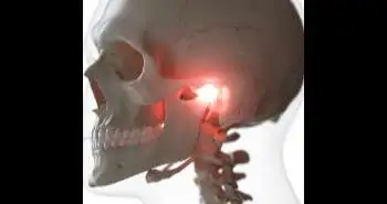
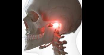
In the present study the efficacy of the conventional TMJ imaging in revealing osseous changes in the mandibular condyle and glenoid fossa in rheumatoid arthritis (RA) and osteoarthritis (OA) patients was evaluated by comparing the findings against CT.
The study was conducted to evaluate the efficacy of the conventional TMJ imaging in showing the osseous changes in the mandibular condyle and glenoid fossa during RA and OA, and the results were compared with the findings of the CT scan. It was found that both radiographic methods are equally efficient in examining the degenerative osseous changes of TMJ in arthritis. But, as the conventional technique does not involve the exposure to unnecessary radiation dosage, it should be preferred for the diagnosis.
In the present study the efficacy of the conventional TMJ imaging in revealing osseous changes in the mandibular condyle and glenoid fossa in rheumatoid arthritis (RA) and osteoarthritis (OA) patients was evaluated by comparing the findings against CT.
A total of 70 patients (40 RA and 30 OA) between age 40-60 years were included in the study and divided into age groups. Then according to clinical history, they were classified as being symptomatic and asymptomatic. Further radiographic examination was carried out. Firstly, the trans-cranial view was obtained (conventional view left and right TMJ), and then the CT Scan was carried out. Then, images were interpreted to report the osseous changes like erosion, flattening, sclerosis and osteophyte formation.
After the comparison of the two radiographic methods, it was observed that both were equally efficient in examining the osseous changed in arthritic patients.
From the analysis it was observed that both the radiographic methods i.e., conventional and CT scan are equally efficacious in examining the degenerative osseous changes of TMJ in arthritis. But, the conventional technique should be preferred as the patient is not exposed to unnecessary radiation dosage.
Curr Rheumatol Rev.
TMJ Arthritis Imaging: Conventional Radiograph Vs CT Scan - Is CT actually needed?
Modgil R et al.


Comments (0)