Categories
Change Password!
Reset Password!


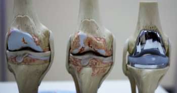
Arthroscopy is “the gold standard” for the diagnosis of knee cartilage lesions.
In this study, the different grades of early degenerative lesions caused due to gonarthrosis were mapped in adults. The knowledge of such lesions were considered helping in knowing the progression of disease and to provide a suitable treatment option.
Arthroscopy is “the gold standard” for the diagnosis of knee cartilage lesions. However, it is invasive and expensive, and displays all the potential complications of an open surgical procedure. Ultra-high-field MRI now offers good opportunities for the indirect assessment of the integrity and structural changes of joint cartilage of the knee. The goal of the present study is to determine the site of early cartilaginous lesions in adults with non-traumatic knee pain.
3-T MRI examinations of 200 asymptomatic knees with standard and three-dimensional double-echo steady-state (3D-DESS) cartilage-specific sequences were prospectively studied for early degenerative lesions of the tibiofemoral joint. Lesions were classified and mapped using the modified Outerbridge and modified International Cartilage Repair Society classifications.
A total of 1437 lesions were detected: 56.1% grade I, 33.5% grade II, 7.2% grade III and 3.3% grade IV. Cartographically, grade I lesions were most common in the anteromedial tibial areas; grade II lesions in the anteromedial L5 femoral areas; and grade III in the centromedial M2 femoral areas.
3-T MRI with standard and 3D-DESS cartilage-specific sequences demonstrated that areas predisposed to early osteoarthritis are the central, lateral and ventromedial tibial plateau, as well as the central and medial femoral condyle.
Br J Radiol.2015 Aug;88(1052):20140542
Mapping tibiofemoral gonarthrosis: an MRI analysis of non-traumatic knee cartilage defects
D S Evangelopoulos et al.


Comments (0)