Categories
Change Password!
Reset Password!


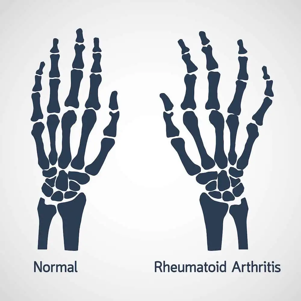
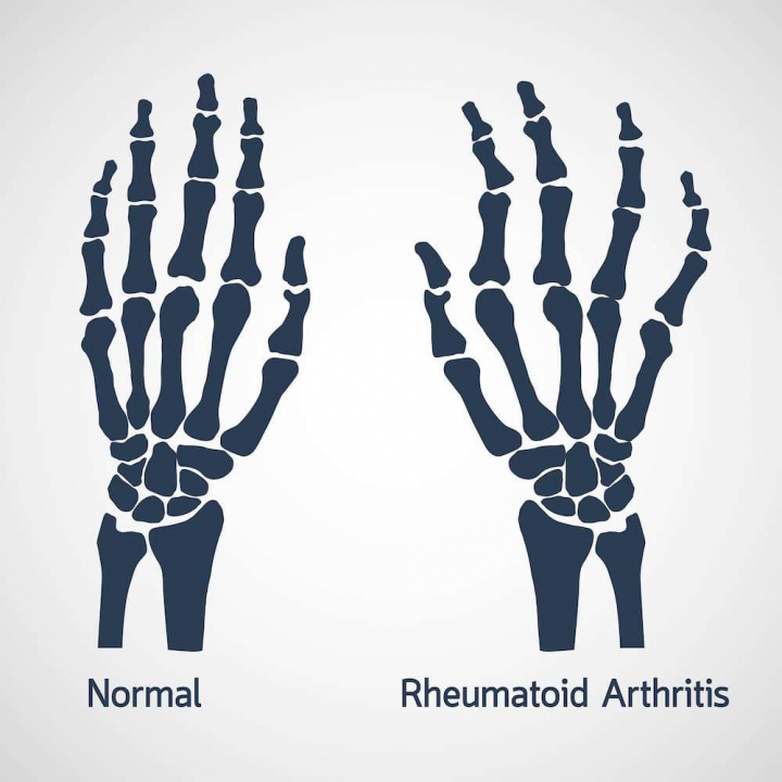
Rheumatoid arthritis (RA) and psoriatic arthritis (PsA) commonly affect the small joints of the wrist and hand.
This study may cater to the in-depth knowledge regarding the pathogenesis of the arthritic process and for non-invasive, objective assessment of rheumatoid arthritis or psoriatic arthritis severity and therapy selection. This research had explored that the high-resolution 18F-FDG PET/CT is indeed a promising tool for the same.
Rheumatoid arthritis (RA) and psoriatic arthritis (PsA) commonly affect the small joints of the wrist and hand. We evaluated the performance of a new, high-resolution extremity positron emission tomography (PET)/CT scanner for characterizing and quantifying pathologies associated with the two arthritides in the wrist and hand joints.
Patients with RA or PsA underwent fluorine- fludeoxyglucose (18F-FDG) PET/CT wrist and hand imaging, respectively, on the high-resolution scanner. Calibrated CT images and co-registered PET images were reconstructed. PET/CT was derived for the radiocarpal and pisiform-triquetral compartments, joints with erosive changes, sites of synovitis or tenosynovitis and the nail bed and were correlated with clinical and MRI findings.
Significantly elevated F-FDG uptake was measured for the radiocarpal and pisiform-triquetral compartments and at sites of bone erosion, synovitis, pannus and oedema, compared with unaffected joints (p < 0.05) in patients with RA, consistent with their clinical findings. In patients with PsA, significantly elevated F-FDG uptake was measured for joints with synovitis compared with unaffected joints (p < 0.05), with patterns of F-FDG uptake along the tendons, at the enthesis and in the nail bed, consistent with tenosynovitis, enthesitis and nail dystrophy, respectively.
High-resolution F-FDG PET/CT imaging of the wrist and hand is feasible in an RA or PsA patient cohort and is capable of providing quantifiable measures of disease activity (synovitis, enthesitis, oedema and bone destruction). High-resolution PET/CT imaging shows promise as a tool for understanding the pathogenesis of the arthritic process and for non-invasive, objective assessment of RA or PsA severity and therapy selection.
Br J Radiol 2016 May 11:20160138
High-resolution 18F-FDG PET/CT for assessing disease activity in rheumatoid and psoriatic arthritis: findings of a prospective pilot study
Chaudhari AJ et al.


Comments (0)