Categories
Change Password!
Reset Password!


This study aimed to distinguish between Ferumoxytol-induced MRI contrast changes with Gadolinium (standard-of-care) in patients symptomatic for osteomyelitis.
Ferumoxytol,
an FDA-approved superparamagnetic iron oxide nanoparticle (SPION) preparation
has been known to be taken up by macrophages in regions of inflammation or
infection, serving as an MRI contrast agent.
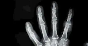
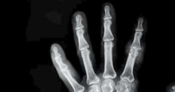
In this study, using ferumoxytol contrasted T 2w images
of patients with osteomyelitis portraying negative augmentation in the
concerned regions. As compared to usual standard of care gadolinium, the images
attained following the ferumoxytol infusion could help in the precise diagnosis
of osteomyelitis.
This study aimed to distinguish between Ferumoxytol-induced MRI contrast changes with Gadolinium (standard-of-care)
in patients symptomatic for osteomyelitis.
Fifteen out of 18 patients
had MRI with both ferumoxytol and gadolinium. As
detected, osteomyelitis was identified in 7 patients, osteomyelitis ruled out
in 5 patients, and 3 patients without a final diagnosis. Mean contrast changes
upon use of ferumoxytol and gadolinium were calculated from relevant lesion
regions. They were then matched with the control regions.
As found, the mean contrast changes i.e. ΔC related with diagnosis of osteomyelitis using Ferumoxytol and T2w imaging sequences were found to be negative i.e. ΔCFe = −2.7 ± 0.7. Whereas for Gadolinium and a T1w imaging sequence, it was positive i.e. ΔCGd = +3.1 ± 1.1 (P < 0.001). Refer the following figure for the same:

As shown in the fig.
Yellow shows patients who did not have osteomyelitis, light purple shows whose diagnosis
was unclear and purple shows the patients who were found to have osteomyelitis.
For both
these agents, the MRI contrast changes correlated with systemic markers of
inflammation (erythrocyte sedimentation rate (ESR)). There was no noteworthy
contrast changes observed in Ferumoxytol-contrasted MRI in patients without
osteomyelitis. The uptake of macrophages in osteomyelitic lesions was no less
than 16X as much iron as the benign bone marrow.
Ferumoxytol-contrasted MRI
seems to be an encouraging method for diagnosing osteomyelitis. Future studies
may be necessary.
Magnetic Resonance Imaging
A comparison of ferumoxytol with gadolinium as contrast agents for the diagnostic magnetic resonance imaging of osteomyelitis
Jens Langsjoen et al.


Comments (0)