Categories
Change Password!
Reset Password!


To assess whether the drop in BMD of the hands (BMD loss), as defined by DXR 3 months after diagnosis, anticipate radiographic joint damage following one and two years within patients with early RA.
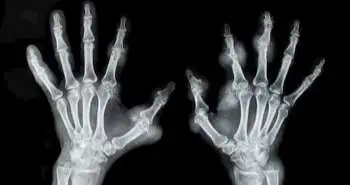
Digital X-ray radiogrammetry (DXR) is computerised analysis technique to estimate peripheral bone mineral density (BMD). Literuture reports that previous DXR-BMD studies have been based on 12-month change, and by that time, conventional X-ray assessments of joint damage are at least as informative about disease progression. This study reports the DXR-BMD loss during the initial 3 months independently predicted radiographic joint damage at 1 year in patients with early RA.
To assess whether the drop in BMD of the hands (BMD loss), as defined by DXR 3 months after diagnosis, anticipate radiographic joint damage following one and two years within patients with early RA.
A total of 167 of these individuals with RA were incorporated in the analysis. Medicine was prescribed under Swedish guidelines, and then a follow up of 2 years was done. Antibodies and rheumatoid factor to cyclic citrullinated peptides were estimated at baseline, and 28-joint Disease Activity Score (DAS28) was evaluated at every visit. Feet and hands radiographs were taken at baseline, three months (hands only), one and two years. The Larsen score assessed baseline and 1-year and 2-year radiographs. Radiographic progression was determined as a difference in Larsen score over the least detectable change. DXR-BMD was estimated at baseline and following three months. The 0.25 and 2.5 mg/cm2/month decline in BMD exhibited a moderate BMD loss and declined up to 2.5 mg/cm2/month described as a severe BMD loss. Multivariate regression was implemented to test the relationship within DXR-BMD loss and radiographic destruction, involving adjustments for possible confounders.
In total, 15% of patients showed severe, and 44% showed moderate DXR-BMD loss at the initial three months. Out of these patients, 19% exhibited radiographic progression at one year and 35% at two years. During multiple regression analyses, a significant relationship was seen between DXR-BMD loss and increase in Larsen score between baseline and one year.
DXR-BMD loss at the first three months autonomously estimated the radiographic joint destruction at one year among individuals with early RA. Thus, DXR-BMD may be a beneficial tool to identify ongoing joint damage and thereby to enhance individualisation of treatment during early RA.
Arthritis Res Ther. 2017 Sep 2;19(1):195
Decrease in bone mineral density during three months after diagnosis of early rheumatoid arthritis measured by digital X-ray radiogrammetry predicts radiographic joint damage after one year
Michael Ziegelasch et al.


Comments (0)