Categories
Change Password!
Reset Password!


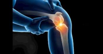
To illustrate the osteophytes (OPs) prevalence identified exclusively by magnetic resonance imaging (MRI) but not by standard X-ray among older adults and to assess longitudinal relationships with knee structural changes.
Radiography fails to detect a large proportion of osteophytes (OP)
which can only be detected by magnetic resonance imaging (MRI). This
population-based cohort study describes that MRI-OPs were associated with
changes in knee structures, and the associations were similar but not as
prominent as those for established-OPs.
To illustrate the osteophytes (OPs) prevalence identified
exclusively by magnetic resonance imaging (MRI) but not by standard X-ray among
older adults and to assess longitudinal relationships with knee structural
changes.
A total of eight hundred thirty-seven participants were
had MRI scans to evaluate knee OPs and additional structures. MRI-detected
early OPs were the OPs which identified only by MRI and established-OPs were
the OPs detected by both X-ray and MRI.
At baseline, the incidence of established-OPs was 10%,
MRI-OPs was 50%, and no-OPs was 40% at total tibiofemoral (TF) compartment. By
adjusting for sex, age, cartilage defects, BMLs, BMI, and/or joint space
narrowing, participants with MRI-OPs as compared to the participants with
no-OPs showed higher risks of increased bone marrow lesions (BMLs) only in
medial TF compartment and cartilage defects in all TF compartments. Further
after adjusting covariates, participants with established-OPs showed higher
risks of increased cartilage defects in total, medial TF compartments and BMLs
in all TF compartments along with higher cartilage volume loss at total and
lateral tibial sites.
MRI-OPs were correlated with changes in knee structures,
and the relationships were alike but not as noticeable as those for
established-OPs. These propose MRI-OPs contribute in knee early-stage
osteoarthritic progression.
Osteoarthritis Cartilage. 2017 Sep 19
Associations between MRI-detected early osteophytes and knee structure in older adults: a population-based cohort study.
Zhu Z et al.


Comments (0)