Categories
Change Password!
Reset Password!


With the technology advancements over the past decade, the treatment options for rheumatoid arthritis (RA) have expanded markedly.
Both dynamic contrast-enhanced MRI (DCE-MRI)
and RA MRI Score (RAMRIS) measures were equally sensitive to treatment effects
with Infliximab.
With the technology advancements over the past decade, the treatment options for rheumatoid arthritis (RA) have expanded markedly. However, the acceptable duration for placebo control has also shortened, with rescue therapy typically offered within 14–16 weeks.
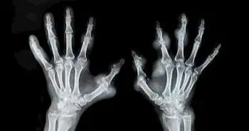
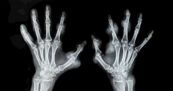
As per the evidences MRI has been more sensitive technique than radiography for detecting joint destruction in RA patients. A method developed by OMERACT (Outcome Measures in Rheumatology) known as RAMRIS is the widely used method for monitoring bone erosion, osteitis and synovitis with MRI in RA. The 9-point cartilage score (CARLOS) is the another most commonly used MRI method for evaluating cartilage loss in RA. Dynamic contrast-enhanced MRI (DCE-MRI) is a quantitative method for assessing synovitis. It is based on the rate and magnitude of enhancement of synovial tissue by intravenously administered gadolinium-based contrast agents (GBCAs). However, it is more difficult to perform than the conventional contrast-enhanced MRI.
The present study by Chan Beals et al. have overcome
these challenges by designing a DCE-MRI technique that simultaneously imaged
the entire wrist and MCP joints. They compared the short-term discriminative
power and sensitivity to change of the volume transfer rate of GBCA from the
blood plasma in synovium (Ktrans) with those of RAMRIS-synovitis in
a randomized, controlled, multicenter trial of infliximab plus methotrexate
(MTX) versus placebo plus MTX in patients with active RA. Both imaging methods
similarly discriminated infliximab treatment from placebo on measures related
to synovial inflammation yet remained stable during placebo treatment. The
RAMRIS and CARLOS methods had additional utility of identifying damage to bone
and cartilage that could be prevented by infliximab. Appropriate MRI
techniques, along with clinical measures of RA activity, should improve the
characterization of drug effects on inflammation and structural damage in RA.
Rationale behind the research:
There are many technical
challenges associated with performing DCE-MRI reproducibly across multiple time
points and multiple clinical sites.
Therefore, the present study was
performed to overcome these challenges by designing a DCE-MRI technique that
simultaneously scans the entire wrist and MCP joints.
Objective:
To compare the scope and the discriminative power of DCE-MRI
with those of semiquantitative MRI scoring for evaluating treatments for RA in
multicenter randomized clinical trials (RCTs).

Study outcome measures:
Primary endpoint: DAS28(CRP) which
is a composite score of the number of tender joints (28 joint count), the
number of swollen joints (28 joint count), patient global assessment of disease
(GADP) on a 100 mm visual analog scale (VAS), and CRP (mg/dL).
Time period: Week 0, 2, 6, and 14

Study Outcomes:
Clinical outcomes:

Figure 2: 2(A) Mean changes from baseline (SE) in DAS28(CRP). 2(B) Mean changes from baseline in Dynamic Contrast Enhanced assessments of the wrist. 2(C) and 2(D) Mean changes from baseline in Dyamic Contrast Enhanced assessments of the metacarpophalengeal joint (MCP) and Enhancing tissue
MRI outcomes:

Figure 3: Baseline vs Week 14 DCE-MRI


Comparison among RA measures:

Safety and tolerability:
In the regulatory guidance, the unresponsive RA
patients recover within 14-16 weeks. With the modern trend towards slowing
structural progression rates, discriminating efficacy reliably with radiography
would be challenging. In many of the RCTs, MRI has been shown to distinguish
suppression of joint damage and inflammation in ≤12 weeks. In the current
study, RAMRIS and CARLOS demonstrated that the bone erosion and cartilage loss
were suppressed with infliximab within 14 weeks.
The results indicate that synovitis and osteitis are
the underlying processes that drive bone erosions and cartilage loss in RA,
which in turn lead to constant pain and physical impairment. In numerous
multi-center RCTs, RAMRIS and CARLOS measures have been used successfully to
demonstrate treatment efficacy. However, none of the studies reported the
successful use of DCE-MRI in a multi-center RCTs. The present study
successfully performed this technique in a multicenter clinical trial using a
knee coil to image both the wrist and MCPs simultaneously. The correlations
demonstrated in this study support the validity of these MRI endpoints as
measures of clinical outcomes in RA. This study suggests that RAMRIS and Ktrans
have similar abilities to discriminate anti-inflammatory treatments. Since
RAMRIS and CARLOS have the broader scope than DCE-MRI and are easier to
implement in multicenter clinical trials, no advantage to recommend the use of
DCE-MRI as applied in this study was reported.
DCE-MRI can be interpreted in a model-free fashion, by measuring the early-enhancement rate and maximal enhancement of the enhancement curves. These empirical measurements are reliable and have been correlated with cellular infiltration and vessel density in the rheumatoid synovium and are responsive to treatment. While somewhat easier to measure, these measurements depend on pulse sequence and machine parameters, rendering comparisons among centers difficult. Because of our interest in methodologies to support clinical testing of RA therapies, we used a compartment model to interpret DCE-MRI. The simple compartment model has one vascular compartment, and one tissue compartment and the measured signal is used to derive the constant of proportionality in the leak of GBCA from capillaries to tissue, Ktrans. Ktrans has been shown to discriminate among patient groups with early arthritis and change with treatment.
In Conclusion, it was demonstrated that both DCE-MRI
and RAMRIS measures of RA inflammation are as sensitive to treatment effects
with infliximab as is the standard clinical measure of RA activity. DAS28(CRP)
identifying impacts as soon as 2 weeks in small numbers of patients.
Furthermore, MRI measures of joint damage (RAMRIS-erosion and CARLOS) can
discriminate treatment effects as early as 14 weeks. In contrast to clinical
tests, however, MRI measures are not vulnerable to placebo effects. Appropriate
MRI techniques, along with clinical measures of RA activity, should improve the
discrimination of drug effects in reducing inflammation and structural damage
in RA.
Appropriate MRI techniques, along with clinical
measures of RA activity, should improve the discrimination of drug effects in
reducing inflammation and structural damage in RA.
PLoS One. 2017 Dec 13; 12(12):e0187397
Magnetic resonance imaging of the hand and wrist in a randomized, double-blind, multicenter, placebo-controlled trial of infliximab for rheumatoid arthritis: Comparison of dynamic contrast enhanced assessments with semi-quantitative scoring
Chan Beals et al.


Comments (0)