Categories
Change Password!
Reset Password!


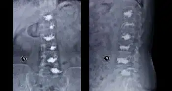
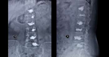
A 60-year-old female patient complained of severe low back pain. She was examined using radio-graphic techniques. After radio-graphic examination of her spine, it was observed that her spine showed degenerative kyphoscoliosis with rotation of the lumbar vertebrae. Her rheumatoid arthritis was controlled but, she still complained about pain. It was also observed that she had no diabetes mellitus.
The most likely diagnosis of this presentation is:
Adult spinal deformity (ASD) often affects a large number of
the elders and its prevalence is increasing day by day. ASD patients experience
greater functional limitations and worse quality of life than the normal
population. Sagittal imbalance is generally correlated with pain. Some authors
also provided the evidence that paravertebral muscles in patients with lumbar
degenerative kyphosis are weak and atrophic with fatty infiltration and
speculated LBP in patients is probably due to fatigue in weak extensor muscles.
Some studies have reported the increased activity in paravertebral muscles in
kyphotic position. In this study, we report the first case of a patient who
presented with painful degenerative kyphoscoliosis and was evaluated with
flourine-18-fluoro-2-deoxy-D-glucose positron-emission tomography/computed
tomography (18F-FDG-PET/CT) preoperatively.
A female
patient with painful degenerative kyphoscoliosis was evaluated with
18F-FDG-PET/CT before surgery. 18F-FDG-PET imaging revealed the uptake of
18FFDG in the paravertebral muscles preoperatively and showed the total lack of
uptake 1-year post-surgery.
Followed by detailed diagnosis, 8F-FDG-PET/CT was performed
immediately prior to the operation at the Department of Endocrine Surgery of
our hospital for the follow-up of thyroid carcinoma. In this, patient fasted
for at least 5h and then FDG was injected in supine position. Her plasma
glucose level was 93 mg/dl. PET scan was done 50 min after the injection. The
maximum standardized uptake value (SUV-max) was 9.7 on the right side and 4.9
on the left side.
As the LBP was not improved by conservative treatment,
surgery was done. Treatment with posterior spinal fusion from Th10 to the ilium
with inter-body fusion and decompression at the level of L3/4, L4/5, and L5/S
was done. Although a spinal orthosis was applied post operatively, proximal
junctional failure with compression fracture of Th11, which caused severe
paraplegia, occurred at 2 months after the primary operation. Therefore, we
performed revision surgery, extending the fusion level to Th2.
This study showed that with the development of surgical
techniques and the improvement in implants, many ASD patients were treated. It
has been reported that vigorous muscle exercise, stress-induced muscle tension,
and activities such as talking or chewing can cause a physiological increase in
the uptake of 18F-FDG in the muscles involved. In this case, the chronically
stretched extensor muscles due to the kyphoscoliotic posture showed a
pathological uptake of 18F-FDG due to the increased muscle activity and that
increased uptake completely disappeared after the appropriate posture was
acquired. Some of the well-known factors causing LBP include intervertebral
disc degeneration, facet joint arthritis, sacro-iliac joint dysfunction, and
paravertebral muscle disorder. In this case, although the patient was receiving
treatment for RA, arthritis of the facet joint or sacro-iliac joint was not
detected on 18F-FDG-PET/CT. On the basis of these findings, LBP in this case
was considered to be caused by chronic fatigue in the paravertebral muscles, which
was visualized with 18F-FDG-PET/CT.
18F-FDG-PET/CT has proven to be an effective treatment in
low back pain which revealed that in the para vertebral muscles was caused by
chronic fatigue. Rheumatoid arthritis was also treated before the surgery. MRI
was also performed which showed the complete disappearance of the increased
uptake in the para vertebral muscles.
Skeletal Radiol..2016 Nov;45(11):1577-81
Potential use of 18F-FDG-PET/CT to visualize hypermetabolism associated with muscle pain in patients with adult spinal deformity: a case report
Taniguchi Y Takahashi M et al.


Comments (0)