Categories
Change Password!
Reset Password!


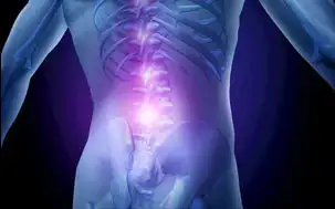
A 34-year-old man of Hispanic origin
visited a clinic complaining of low back pain radiating into left anterolateral
thigh that he was experiencing for four months. The severity of pain was
varying and often worsened on walking and with rigorous activity. The patient
also reported a new complaint of mild left anterior hip stiffness. There was no
history of trauma or any accident. Intervertebral disc protrusions at the L4/5
and L5/S1 levels were observed in the lumbar spine MRI did at an outside
institution.
The most likely diagnosis
of this presentation is
Low back pain (LBP) is the most common
ailment affecting work performances, well-being and overall quality of life.
The pain may range from acute, or subacute to chronic and may be described as
an aching, well defined, burning, stabbing, sharp, vague or dull. Infection,
ankylosing spondylitis, tumour, fracture, inflammatory process, osteoporosis,
and radicular syndrome are likely to be the factors causing back pain.
Depression, ageing, occupational posture and obesity are the established risk
factors. The current treatment regimen for LBP involves pain-relieving
medicines, rehabilitation therapies and surgical approach as a last resort. The
exact underlying cause of LBP is often challenging to identify, thereby making
the diagnosis and treatment challenging.
Nontraumatic osteonecrosis of the femoral head (ONFH) is caused by the degradation of bone cells resulting in subsequent osteoarthritis. It mainly affects elderly patients aged 30 to 40 years. Contributing causes of ONFH may include corticosteroids, alcohol abuse, history of trauma, hemoglobinopathy, Gaucher disease, coagulopathies, and other severe ailments. Osteonecrosis (ON) of the hip is characterized by the insufficient nutrient blood supply to the femoral head causing structural failure of the cortical surface. LBP can also result from concurrent conditions.
Moreover, patients with ON report
concomitant pain in the low back, buttock, groin, thigh, and knee. Studies have
reported the examples of LBP remotely generated from the hip osteoarthritis in
patients who underwent total hip arthroplasty and experienced reduction in hip
and LBP symptoms. Hip ailments as an underlying cause of LBP may be attributed
to the functional interdependence of related regions and thus called as a
hip-spine syndrome.
The patient presented with low back pain was diagnosed
with bilateral idiopathic osteonecrosis of the femoral head, suggesting
consideration of differential diagnosis. The patient recovered after THA of the
left hip.
On careful physical examination, thoracolumbar spine
showed decreased range of motion. At the end range of flexion, increased spinal
pain was noted. Lumbar spine movement was decreased, and the musculature was
sensitive to touch at all points. No motor or sensory deformities were
observed. Spinal instability was revealed from the prone instability test and
Kemp’s test. Positive modified Thomas test revealed hip flexor hypertonicity.
Based on this, the patient was diagnosed with dysfunction of lumbar segments,
myofascial pain syndrome and dislocation of lumbar intervertebral discs without
myelopathy. Accordingly, treatment was initiated. But due to continuous
distress on the left side of the limp, the patient had to undergo re-examination.
While evaluating the left hip, a positive C sign, in which the patient “cups”
the anterior hip with their thumb and forefinger as to make the letter C, and
considerable pain in the anterior hip during FABERE (Patrick’s), hip
impingement, McCarthy and modified Thomas tests was observed. Left hip standard
X-rays were carried out to eliminate hip impingement; showed signs of
osteonecrosis, i.e. ill-defined sclerosis and collapse of the articular surface
with the fragmentation of the femoral head. Bilateral hip MRI examination
confirmed the osteonecrosis of the left femoral head.
Chiropractic treatments, including
palliative care, manipulative therapies, and exercises, were employed to
relieve the pain. Exercise programs that included McKenzie repetitive extension
exercises and computerized traction/decompression therapy were given to the
patient two times a week for three weeks. The patient experienced relief in low
back pain. The patient was evaluated for the need for surgical intervention and
was finally treated with bilateral total hip arthroplasty (THA).
In the present case, a detailed diagnosis was not
suggestive of a primary source of LBP originating from low back structures.
Radiological studies also did not indicate underlying pathology in the lumbar
spine. Assessment of the hip was mildly suggestive of several chronic ailments,
including femoroacetabular impingement and osteoarthritis. Differentiating low
back from hip pathology can be challenging due to overlapping pain referral
patterns. It is difficult to distinguish pain originating from low back to pain
originating from the hip in older patients due to degenerative changes. A brief
assessment of a history and physical evaluation of the low back and hip regions
may help in accurate and timely diagnosis.
The hip MRI confirmed ONFH in the patient under consideration. At preliminary diagnosis, ONFH can be bilateral in up to 60% of cases. The risk of further development of ONFH is very less if MRI of contralateral hip shows normal findings at preliminary diagnosis. The patient may show unilateral symptoms even in the presence of bilateral ONFH. The symptoms include significant hip pain, mild anterior hip stiffness, gait disturbances and limited range of motion.
MRI should be considered as the most sensitive tool for
detecting ONFH as standard X-rays depicts ONFH pathological signs (patchy
sclerosis, fragmentation of femoral head) at a very late stage of the disease.
At the early stage, ONFH can be treated by core decompression with implantation
of a tantalum rod that provides structural support by acting as a buttress for
the subchondral bone and encouraging bone ingrowth around the rod. However,
sometimes patients operated on with decompression may experience more severe pain
and restricted walking and had to undergo THA. Bone marrow mesenchymal stem
cells (BMMSCs) have the potential of self-proliferation and multi-potential
differentiation, therefore, can be induced to undergo osteogenesis. Thus,
effective alternate therapy can be introduced.
While using MRI, the contralateral side not showing prominent symptoms should not be considered.
ONFH even the absence of risk factors should be suspected in older adults with significant hip pain or discomfort, not responding well to the treatment.
Differential diagnosis for the hip pain followed by diagnostic imaging could help in early recognition and timely treatment of the disease.
J Chiropr Med. 2014 Sep; 13(3): 196–202.
Bilateral Idiopathic Osteonecrosis of the Femoral Head: A Case Report With an Emphasis on Differential Diagnosis, Imaging, and Treatment
Patrick J. Battaglia et al.


Comments (2)