Categories
Change Password!
Reset Password!


A new nomogram was found to be
valuable to predict the radiographic advancement of osteoarthritis and enhance
the predictive capability for clinicians and researchers.
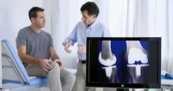
The data from Foundation for the National Institutes of Health (FNIH) osteoarthritis biomarkers consortium project revealed that nomograms based on three-dimensional (3D)-magnetic resonance imaging (MRI) bone shape possess excellent predictive value for mild to moderate osteoarthritis advancement. The bone shape alteration at 24 months exhibits superior predictive value compared to bone shape at baseline.
Researchers undertook this study for developing and validating a new nomogram for predicting advancement of osteoarthritis on the basis of 3D-MRI bone shape and alteration in bone shape during 24 months. From the baseline, the minimum radiographic narrowing of medial tibiofemoral joint space of ≥ 0.7 mm at 24, 36, or 48 months was defined as radiographic advancement. There were 303 knees without radiographic advancement and 297 knees with radiographic advancement.
The bone shapes of femur, patella, tibia were determined by 3D-MRI at baseline and at twenty-four months. Separate establishments of 2 nomograms were done with the help of multivariate logistic regression assessment employing clinical risk factors, bone shape alteration at twenty-four months (nomogram Δ24), or bone shape at baseline (nomogram 0).
For assessing the nomograms, selection of calibration, usefulness, and discrimination was made. Profound differences were reported between the groups in baseline Kellgren-Lawrence (KL) grade, sex, age, and shape of patella, tibia, and femur. The areas under the curve (AUC) and accuracy of nomograms at baseline and twenty-four months are shown in Table 1:

Both the nomograms exhibited good
calibration. According to decision curve analysis, nomogram Δ24 revealed better
clinical utility than nomogram 0 when the risk threshold was noted to range
from 0.04 to 0.86. Thus, this study highlighted the close association between
bone shape alteration and the progression of osteoarthritis.
BMC Musculoskeletal Disorders
Novel nomogram for predicting the progression of osteoarthritis based on 3D-MRI bone shape: data from the FNIH osteoarthritis biomarkers consortium
Yingwei Sun et al.


Comments (0)