Categories
Change Password!
Reset Password!


In the absence of specific microscopic techniques, ultrasonography that identifies the double contour sign, intra-articular aggregates, and/or tophi could be utilized for diagnosing gout.
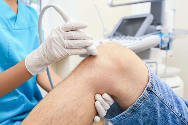
Gout refers to a condition in which needle-like uric acid crystals deposit in the joints. The deposition of uric acid can lead to inflammation, pain, and redness in joints described as acute arthritis. The diagnosis of gout can involve a painful aspiration of the synovial fluid. So, it is important to explore other non-invasive alternatives which could aid in diagnosing gouty arthritis.
Nuttaya Pattamapaspong and colleagues conducted a study to assess the usefulness of ultrasonographic (US) characteristics of crystal deposition for detecting gout in patients presenting with acute arthritis. A total of 89 patients with acute arthritis were enrolled, and ultrasound scanning of the most inflamed joint was executed on them. Two radiologists separately analyzed the ultrasound images, and in case of any variation in the four ultrasound features, consent was taken from a third radiologist. The gold standards for diagnosing gouty arthritis are arthrocentesis and crystal analysis using compensated polarized light microscopy of aspirates.
Results indicated that fifty-three (60%) patients had gout, whereas the remaining thirty-six (40%) had non-gout arthritis. Mean serum uric acid level was 7.1 mg/dl and 4.7 mg/dl in patients with gout and non-gout arthritis respectively. Changes in three US features, the double contour sign (42 vs. 8%), intra-articular aggregates (58 vs. 8%), and tophi (40 vs. 0%) were observed (p < 0.001) between patients with gout and non-gout arthritis. There were no clinically important differences found in detecting intra-tendinous aggregates (32 vs. 17%, p = 0.14). The sensitivity and specificity of above three US features were estimated and found to be 42 and 92% for double contour signs; 58 and 92% for intra-articular aggregates; and 40 and 100% for tophi respectively. For these three US features, the positive predictive values varied from 88 to 100%, while the negative predictive values varied from 52 to 60%.
It was concluded that the above described US features; double contour sign, intra-articular aggregates, and tophi could be a useful addition to the diagnosis of acute gout, mainly when prevalence rate is high and specialized microscopic techniques are not available.
Skeletal Radiology
Value of ultrasonography in the diagnosis of gout in patients presenting with acute arthritis
Pattamapaspong et al.


Comments (0)