Categories
Change Password!
Reset Password!


The conventional
radiographs can now be replaced with structural lesions on MRI to classify
patients as per the ASAS axSpA criteria.
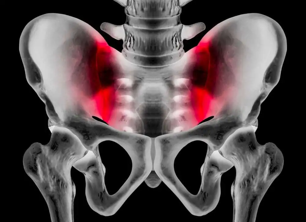
Axial Spondyloarthritis, an inflammatory arthritis condition of spine and
pelvic joints, causes severe pain and muscle stiffness. To decrease severe pain
and unnecessary therapeutic procedures, early diagnosis and therapeutic
interventions are very important. This early diagnosis can be achieved by means
of MRI studies. In radiographs, inflammation of spine and structural
lesions of sacroiliac joints can visible before starting of structural damage.
Therefore, to
serve the case, a study has been conducted to check the reliability of
structural lesions seen on MRI for classification of patients in accordance
with (ASAS) axial SpondyloArthritis (axSpA) criteria in DESIR cohort.
A total of 582
patients were participating in the study. The patients who were under the age
of 50 and showed symptoms that last for 3 months to 3 years were selected for
the study. The diagnostic studies such as MRI T1-w images (structural lesions,
MRI-SI-s) MRI STIR (inflammation, MRI-SI) and sacroiliac joints radiographs
(X-SI) were scored by two well – calibrated readers. Further, the MRI-SI and
X-SI difference was adjudged by a third reader. For the study, formerly
proposed cut-offs of positive MRI-SI-s with more than 5 erosions & fatty
lesions (E/FL ≥5) were taken.
The analysis was
done and out of 582 patients, only 418 fulfilled the ASAS axSpA criteria. Out
of which 127 patients were evaluated as modified New York (mNY) positive and
134 (according to reader 1) & 75 (according to reader 2) were as MRI-SI-s
positive. The mNY and MRI-SI-s agreement was in the balance with each other
(reader1,k:0.39; reader2,k:0.44). Therefore, if E/FL≥5 cut-off was used rather
than mNY, no change in classification was seen in 478 patients of reader 1 and
469 of reader 2. Furthermore, in condition of mNY replacement, where only
MRI-SI-s was performed, 12 patients of reader 1 & 10 of reader 2 might not
classify as axial SpondyloArthritis. On the other hand, in both scenarios, 3
patients of reader 1 & 6 of reader 2 might be additionally classified as axial
SpondyloArthritis. The same kind of results was noticed for E≥3 and FL≥3
cut-offs.
The whole analysis reach to a conclusion that the structural lesions of sacroiliac joints on MRI are reliable for axSpA diagnosis either as additionally or as a substitute for radiographs in the ASAS axSpA classification.
Annals of Rheumatic Diseases
Can we use structural lesions seen on MRI of the sacroiliac joints reliably for the classification of patients according to the ASAS axial spondyloarthritis criteria? Data from the DESIR cohort
Pauline A C Bakker et al.


Comments (0)