Categories
Change Password!
Reset Password!


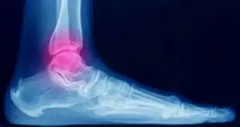
Ultrasound is as functional as conventional X-ray images in diagnosing ankle sprain-related fractures.
Ultrasound is at least as useful as conventional X-ray images in the diagnosis of ankle sprain-related fractures, notwithstanding age or sex and could, in fact, be better, as mentioned in the 'European Journal of Trauma and Emergency Surgery'.
In traumatology, Ankle torque is the most commonly encountered injury mechanism. It mostly develops as an ankle sprain and often leads to an ankle fracture. The signs and symptoms are very unspecific on the differential diagnosis, and the conventional radiographs must be obtained to rectify it. Ferràs-Tarragó J and investigators compared the ultrasound and standard X-ray images values found in ankle sprain-related fracture diagnoses. A total of 52 patients with ankle torque were considered for this 3-month prospective study. An ultrasound diagnosis was performed by the first researcher at their arrival to the emergency department (ED), mostly consisting of a longitudinal section of the fibula, tibia, and V metatarsal. A blinded independent investigator then carried out the usual diagnosis protocol via the traditional radiographs. The third independent investigator assessed the results when the required number was obtained. A Chi-squared test helped to contrast the outcomes between sensitivity, specificity, positive predictive value (PPV), and negative predictive value (NPV) distinguishing a non-inferiority hypothesis of our protocol against the standard X-ray images screening. On the detection of ankle torque-related fractures, echography portrayed to be at least as good as the standard radiographs. Nearly 8% of fractures were misdiagnosed with plain radiography in these patients, which is as per the lower limit found in the bibliography. In the first ultrasound assessment, all the false negatives on radiographs were true positives. The average time for the ultrasound protocol was 42 seconds.
The investigators concluded," The use of echography could decrease the number of radiography performed when diagnosing these kinds of fractures, hence reducing the amount of radiation exposure and expediting the diagnostic process and also the "in situ diagnosis".
European Journal of Trauma and Emergency Surgery
Ankle torque-related fractures and its echo-fast diagnosis protocol
Ferràs-Tarragó J et al.


Comments (0)